

Confocal Microscopy (CFM) | Free-Beam Setup
Confocal microscopy is the most common instrumental technique in quantum optics to investigate semiconductor quantum dots, single molecules, color centers in diamond, photonic crystals, nanowires, quantum wells, 2D layered materials (graphene, chalcogenides) and many more. Often, the quantum nature of light emission requires cryogenic temperatures for the samples under investigation. Low temperatures also result in sharper spectral lines due to decreased thermal broadening, as well as in stronger optical signals, since the quantum efficiency is improved due to less scattering and less non-radiative recombinations. In addition, high magnetic fields can be used to extract information about the energy levels.
A confocal microscope is able to increase the optical resolution compared to conventional microscopes by rejecting out-of focus light (see schematics below):
1. The sample is illuminated with a focused beam, which restricts the excitation to a small region on the sample both in lateral as well as
vertical direction.
2. The use of a blocking pinhole in the conjugate plane eliminates any out-of-focus information in the collection path.
As in most of attocube’s low temperature microscopes, an image is acquired by scanning the sample, while the probe, in this case the objective, remains fixed. This technique enables diffraction limited lateral optical resolution, as well as 3D imaging with very good vertical resolution by scanning several thin sections at different focal planes through the specimen. These advantages come at the price of lower collected intensity, and hence longer integration times.
The shown schematics is usually referred to as ‘free-beam’ configuration, which enables to add a number of filters to either the excitation, the collection or the combined beam path. This enables sophisticated spectroscopic as well as imaging techniques such as resonant fluorescence, Raman spectroscopy or polarization analysis.
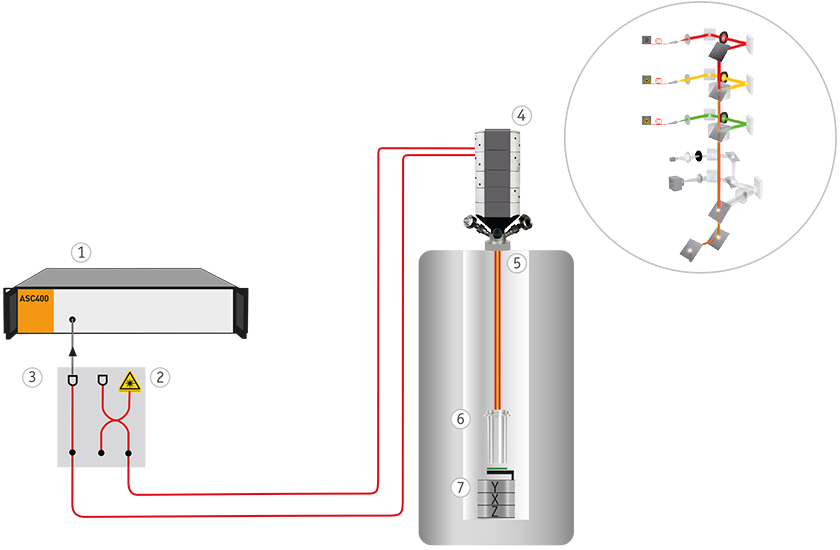
- ASC400 scan controller
- LDM600 laser source
- LDM600 2nd detector
- attoCFM I external optics head incl. optional filters
- Optical top window
- Free-beam based cryogenic LT-APO objective
- Sample with xyz positioners and optional xy scanner