Ultimate Thermal Stability and Ultra-Low Drift
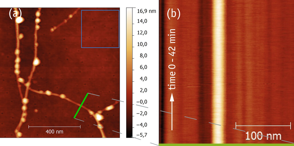
In order to characterize both the low-frequency drift of the atomic force miroscope unit of the attoCSFM with respect to the sample, a carbon nanotube (CNT) was imaged (a). By scanning the same line across the CNT (green line in overview image) 500 subsequent times within 42 minutes (b), a line-to-line position jitter below 1 nm and a long-term drift of less than 3 nm were observed (c), demonstrating the outstanding thermal and mechanical stability of the attoCSFM. After several hours of thermalization, drifts below 1 nm/h can be achieved (d). The part of drift due to scanner creep is constantly monitored interferometrically and can therefore be corrected.
(attocube application labs, 2012; sample courtesy of A. Hartschuh, LMU Munich, Germany)
This measurement was realized with the Combined Atomic Force & Confocal Microscope.


Room temperature platform for ODMR
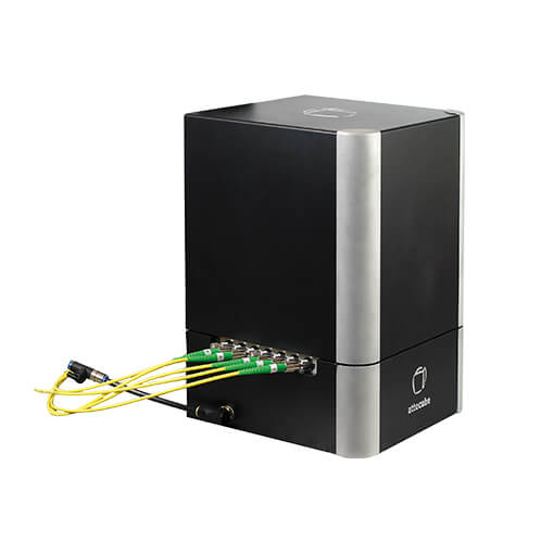
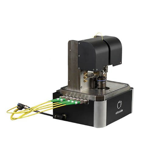
After decades of evolution in magnetic imaging, combining the sensitivity needed to detect single electron or nuclear spins with a spatial resolution of a few nanometers may soon become current state-of-the-art instrumentation: nitrogen vacancy (NV) nanomagnetometry based on the principle of optically detected magnetic resonance (ODMR) is commonly considered the most promising candidate to achieve this goal. While there is significant scientific activity to both reliably prepare appropriate nanodiamonds with tailored NV center characteristics and to attach them onto AFM tips, attocube is complementing these efforts by providing an ideal platform for ODMR: the attoCSFM combines a high-NA confocal microscope for optical detection in transmission with a cantilever based atomic force microscope, completely built from non-magnetic materials.
The sample environment can be adjusted from ambient pressure to low vacuum (≈1 mbar), providing integrated temperature control and subsequent drifts to less than 10 nm/h. Precise relative positioning of the sample, AFM tip, high NA objective, and an optional permanent magnet is enabled via 10 degrees of freedom provided by nanopositioning stages and scanners (positioning ranges 15 mm x 15 mm x 15 mm, scan ranges 20 µm x 20 µm x 7 µm).
This measurement was realized with the Combined Atomic Force & Confocal Microscope.


