

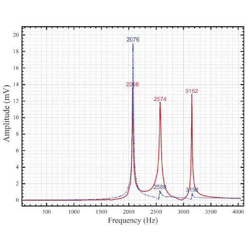
Resonance Frequency of an ANSxy50 made of titanium
attocube scanners, especially the ones made for cryogenic and magnetic environments are often implemented in highly sensitive scanning probe microscopes. One of the major issues in such setups is stability and vibrational stiffness. For such an application, the group of Bert Voigtländer from the institute of Quantum Nanoscience at Forschungszentrum Jülich (Jülich, Germany) detected the resonance frequencies of a standard ANSxy50 positioner made of titanium. The top plate of the scanner was removed for their purpose and the device was fixed on a support. The scanner body was excited with an external piezoelectric element while the response voltage (due to the piezoelectric effect) of the integrated piezo elements detected the resulting vibrations of the scanner. The measurements showed clear resonance peaks at frequencies in the kHz regime, namely 2.1 kHz (1st mode), 2.6 kHz (2nd mode) and 3.2 kHz (3rd mode), showing the high stability of this attocube scanner. Comparing the results with simulations of the resonances idetified these with complex modes with dominant vibrating directions in z (1st mode), and in perpendicular modes in the x-y plane (2nd and 3rd mode). After this successful evaluation, the Jülich group will use these scanners, which combine high stiffness with a large scan range, in a low temperature four-tip scanning probe microscope.
This measurement was realized with the ANSxy50/LT xy-Scanner made from Titanium.
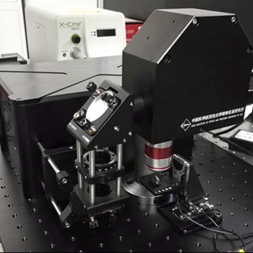
Life Science: 3D Imaging System Showing Natural Colour
Interesting features in life science and biology often span several orders of magnitude in size. A complete optical study requires to resolve structures from sub-micrometer to centimeter, thus high resolution is a fundamental need. In addition, fast acquisition times are demand, as any movement during the image-taking process will cause blurr.
The group of Prof. Ming Lei at the State Key Laboratory of Transient Optics and Photonics (Xi'an Institute of Optics and Precision Mechanics, Chinese Academy of Sciences, Xi’an, China) developed such a 3D microscope with a lateral resolution of 580 nm.
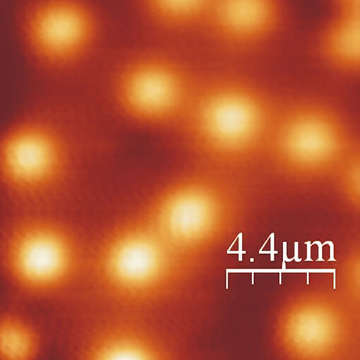
The possibility to obtain full colour 3D images and 3D morphological data in the size range of typical insects opens the door for many different types of entomological investigations. Three attocube ECS positioners actuate a microscopic stage to move and align the sample in all three dimensions. The positioners are perfectly suited for such an application as they provide a travel range of at least 20 mm with a typical step size of less than 100 nm.
This measurement was realized with the ECSx3030/Al/NUM/RT.
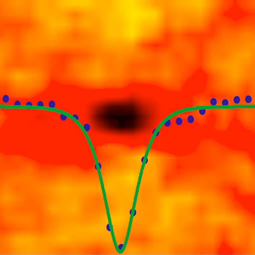
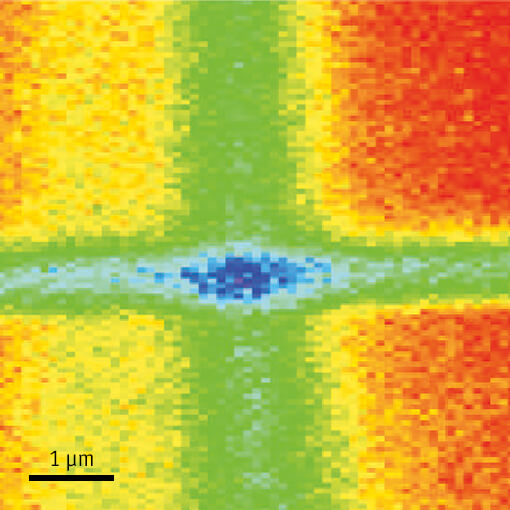
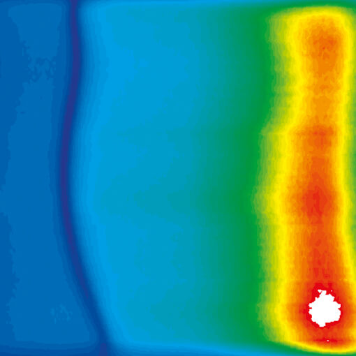
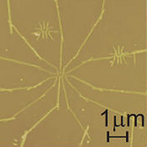
Sample positioning in scanning transmission x-ray microscope at SLAC
We use attocube linear stages with optical and resistive encoders for positioning of samples relative to high resolution x-ray optics in a scanning transmission x-ray microscope. The picture shows a magnetic excitation called “soliton” that is the result of a spin polarized current injected into a thin ferromagnetic Co/Pt multilayer. The injected spin causes a localized oscillation of the magnetization at the spot where the current is injected, here a nano contact with a 150 nm diameter in the center of the picture. Time resolved scanning transmission x-ray microscopy using magnetic circular dichroism as a contrast mechanism is used to obtain an image of the magnetic soliton.
(Dr. Hendrik Ohldag, Stanford Synchrotron Radiation Laboratory, SLAC National Accelerator Laboratory.)
This measurement was realized with the ANPx101/UHV - linear x-nanopositioner, and the ANPz102/UHV - linear z-nanopositioner.
Characterizing a scanning fluorescence X-ray microscope
When developing an X-Ray microscope capable of nm resolution, careful design is a must. Thermal and mechanical stability of the components and assemblies has to be followed throughout the process. The FPS shows superior performance regarding its outstanding stability and its capability of measuring sub-nm displacements. The senor has a better than 1.25 nm stability over 40 hours, and a better then 300 pm resolution at 100 Hz bandwidth in a controlled environment. The FPS is therefore the ideal supplement for the mechanical control of all components used in the described X-Ray microscope setup achieving a resolution in the order of 40 nm, while the stability is below 45 nm over the entire time needed for data collection.
This measurement was realized with the Displacement Measuring Interferometer.
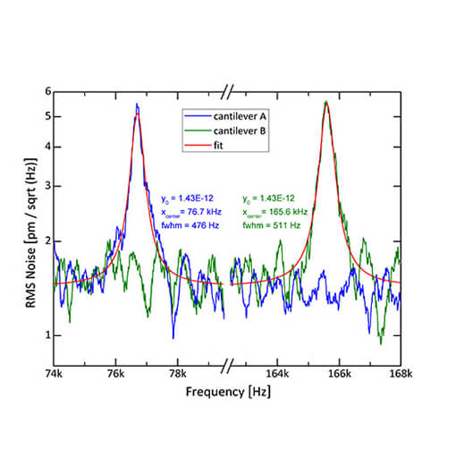
Measuring Brownian Motion of Comercial Micro-Cantilevers
Frequency analysis is a standard method to study demanding applications and measure the frequency dependent mechanical motion of e.g. micro- or nanomechanical systems (MEMS / NEMS). Measuring vibrations with amplitudes of only few picometers is very challenging, exceeding the capabilities of other commercially available measurement techniques.
To demonstrate that attocube’s fiber-based Displacement Sensor (IDS) not only has high-resolution but also a very low noise floor, we measured the resonant vibrations of micro-sized cantilevers which were excited only by their thermal energy at ambient conditions.
This measurement was realized with the Displacement Measuring Interferometer, and the .
Performance Test of the ANPz30/LT at 35 mK and 15 Tesla
The precise performance of nanopositioning elements is of great importance in order to realize instrumental setups which work reliably under extreme environmental conditions. Although attocube systems’ positioners have been tested at low temperatures down to 10 mK and at high magnetic fields up to 28 Tesla, their successful performance has never been demonstrated when both environmental conditions were simultaneously applied. A real challenge, furthermore, is to carry out such a test in a 3He/4He environment due to the fact that 3He carries a magnetic spin which becomes polarized in magnetic fields. This influence on the positioner’s operation was investigated for the first time in this application.
This measurement was realized with the ANPz30/LT - linear z-nanopositioner.
Magnetic Resonance Imaging of Nanoscale Virus at 300 mK
attocube’s ANPx51 positioners were used in an MRFM setup with the task to precisely and reliably position a magnetic tip and a copper nanowire to close proximity of an ultra-sensitive cantilever. The MRFM setup was applied to investigate and reconstruct the 1H spin distribution of Tobacco Mosaic Virus particles, representing a 100-million fold improvement in volume resolution over conventional MRI.
This measurement was realized with the ANPx51/LT - linear x-nanopositioner.
Photoluminescence measurements in fields up to 28 T
The attocube systems positioners ANPxyz100/LT have been used in a setup for optical measurements in liquid 4He temperature and magnetic fields up to 28 T at the Grenoble High Magnetic Field Laboratory. In the setup laser excitation is delivered using a single-mode fiber and is focused onto the sample with two microlenses. A multimode fiber is used for photoluminescence (PL) collection.
This measurement was realized with the ANPx101/LT - linear x-nanopositioner, and the ANPz102/RES/LT - linear z-nanopositioner.
Scanning Hall Probe Microscopy at 300 mK with ANP positioners
The magnetic properties of superconducting and ferromagnetic materials at ultra-low temperatures represent some of the most interesting contemporary problems in condensed matter physics. These properties are typically investigated using a magnetic force microscope or a scanning Hall probe microscope (SHPM). In this note, we report on a self-built SHPM capable of working at temperatures as low as 300 mK and magnetic fields of up to 10 T, while still having sub-micron lateral spatial resolution.
This measurement was realized with the ANPx101/LT - linear x-nanopositioner, and the ANPz102/RES/LT - linear z-nanopositioner.
Mapping and Manipulation of Leakage Currents in a Nanostructure
In this application note, attocube’s smallest titanium positioners (ANPx51/RES and ANPz51/RES) are used as part of an atomic force microscope (AFM) inside a Janis ³He cryostat with a base temperature of 280 mK. The setup is a combined low temperature AFM and scanning tunneling microscope (STM), which is emploied to carry out scanning gate microscopy experiments on various nanostructures. In these measurements the positioners are used to move the metallic tip directly above the nanostructure predominantly at 4.2 K but also as low as 280 mK.
This measurement was realized with the ANPx101/RES/LT - linear x-nanopositioner, and the ANPz102/RES/LT - linear z-nanopositioner.
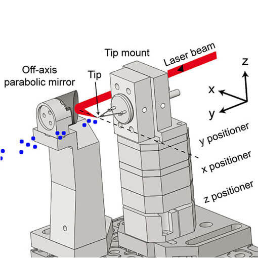
Controlling Ultra-Fast Electron Emission
The dynamics of electrons emitted from a sharp tungsten tip triggered by femtosecond laser pulses have been investigated. The setup shown to the left is situated in an UHV chamber at p = E-10 mbar pressure. A xyz positioning stack enables precision alignment of the tip. Photoelectron spectra are recorded while the phase between carrier wave and intensity envelope is varied in small steps. The lower figure shows two electron spectra, recorded with a phase difference of 180°. In a), pronounced peaks are visible caused by interference of two electron wave packets emitted during subsequent optical cycles. In b), no peak structure is visible; only one electron wave packet contributes. This energy domain effect allows conclusions about the time dynamics of the electrons. By shaping the laser electric field with the carrier-envelope phase, the dynamics of the electrons can be controlled with attosecond precision. The presented system enables control over photoelectrons from a metal tip in space (nanometer scale) and time (attosecond scale).
(The data was kindly provided by M. Krüger, M. Schenk, and P. Hommelhoff, Max Planck Institute of Quantum Optics, Garching, Germany.)
This measurement was realized with the ANPx101/UHV - linear x-nanopositioner, and the ANPz102/UHV - linear z-nanopositioner.