
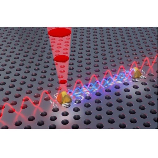
Interacting single-photon emitters
Quantum photonics research focuses on miniaturized devices that transmit quantum states of light. The quantum states which are the most sought after are entangled states, because they are necessary for realization of quantum communication and quantum computing. The team lead by Peter Lodahl (Niels Bohr Institute, University of Copenhagen, Denmark) has achieved a major breakthrough – interaction between two single-photon emitters (SPEs). They have achieved interaction by embedding SPEs in a photonic crystal waveguide, that extended the range of the dipole-dipole coupling, which would otherwise be very short-ranged, i.e. substantially smaller than the corresponding wavelength. They have measured collective response of two interacting emitters and demonstrated ability to control them simultaneously. In this study they have been using an attoCFM I confocal microscope in an attoDRY1000 cryostat, where they placed their quantum photonics chip. Their achievement represents reaching a long-awaited milestone on the quantum roadmap, and is essential for scaling up of quantum computing and quantum communication technology.
This measurement was realized with the attoDRY1000, and the attoCFM I.
Further reading:
A. Tiranov et al., Science 379, 389 (2023)
Excitons in TMD for valleytronics
Semiconducting single-layer transition metal dichalcogenides (TMDs) have proven to be well suited for studying interplay between charge, spin and lattice, which is of interest for both fundamental physics and development of future applications. The broken inversion symmetry in TMDs and strong spin-orbit coupling lead to the spin-valley coupling of band edge electrons. In turn, a collaboration led by Xiaodong Xu (University of Washington, USA) has observed a new series of low-energy excitons, strongly coupled to phonons, in the photoluminescence (PL) spectrum of ultraclean single-layer WSe2, using an attoCFM I microscope in an attoDRY2100 cryostat. Their findings open a gate towards exploring collective phenomena in quantum optics using intrinsic donor-bound excitons in 2D materials and towards designing optical spin quantum memory.
This measurement was realized with the attoCFM I, and the attoDRY2100.
Further reading:
P. Rivera et al., Nature Commun. 12, 871 (2021)
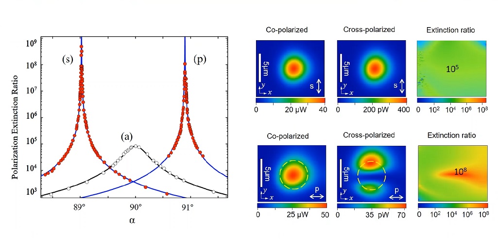
Extreme cross-polarization extinction
Confocal microscopy has proven to be an essential tool for studying light-matter interactions via resonant fluorescence experiments on the sub-micron scale. However, the exact mechanism behind the surprisingly strong suppression of laser light in such a configuration remained elusive.
In the framework of a PhD thesis conducted at attocube and financed through the EU’s Horizon 2020 network 4PHOTON, its origin could be attributed to the Imbert-Fedorov shift, a subtle sideways shift of a polarized beam of light when it is reflected off a smooth surface. For the precise rotation of the polarizers, an attocube piezo stepper rotator was used, whose accuracy of 15 µRad steps is essential to achieving such extreme extinction values.
Our work opens the way to a methodical design of sensitive laser resonant fluorescence microscopes with extreme background extinction, for a broad range of applications in quantum optics and solid-state physics, as well as for measuring material optical properties.
This measurement was realized with the attoCFM I.
Further reading:
M. Benelajla et al., Phys. Rev. X 11, 021007 (2021)
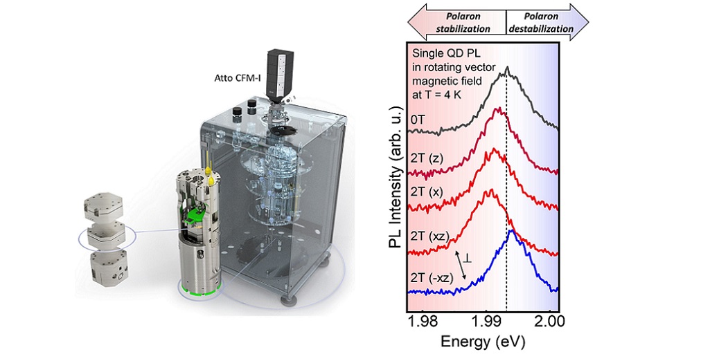
Anisotropic formation of exciton magnetic polarons in colloidal quantum dot
Colloidal nanocrystal quantum dots are - due to their reproducible and scalable fabrication process - promising candidates for many industrial applications like solar cells, LEDs and displays. Doping semiconducting colloidal nanocrystals with magnetic ions can lead to extraordinary effects, such as light-induced magnetization - aka exciton magnetic polaron (EMP). The group of Gerd Bacher (University of Duisburg-Essen, Germany) carried out temperature- and magnetic-field-dependent photoluminescence measurements on single colloidal Mn2+:CdSe/CdS quantum dots, thus shedding light on the fundamental processes that lead to EMP formation. The measurements were realized with attoCFM I in attoDRY1000 with 3D magnetic field. These findings open a new path for controlling the orientation of light-induced magnetism.
This measurement was realized with the attoDRY1000, and the attoCFM I.
Further reading:
S. Lorenz et al., Nano Lett. 20, 1896 (2020)

Single photon sources on the way to QIP
Single-photon sources will be a fundamental building block for future quantum information devices. For advanced implementations, the photon sources must emit simultaneously high efficiency and indistinguishability photons. On the way towards an optimal solid-state based single-photon source, the group of Chaoyang Lu and Jian-Wei Pan at the University of Science and Technology of China (Shanghai, CN) presented a background-free method (two-color excitation) for generating spectrally isolated indistinguishable photons [1] and polarized single photons from elliptical micro-pillars. The optical measurements were performed using an attoCFM I cooled by a closed cycle attoDRY2100 cryostat. With their measurement method they demonstrated a state-of-the-art polarized single-photon efficiency of up to 60% and an indistinguishability of up to 0.975 for the micro pillar device [2], which allowed them to perform the first 20 photon experiment leading toward quantum supremacy [3].
This measurement was realized with the attoDRY1000, and the attoCFM I.
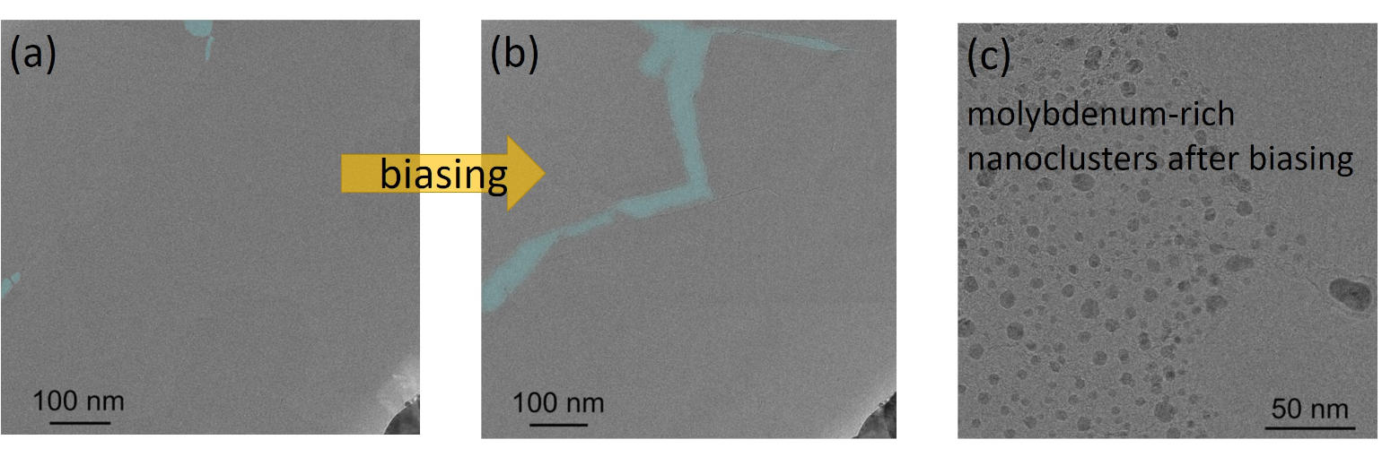
In-situ electrical biasing technique in MoS2
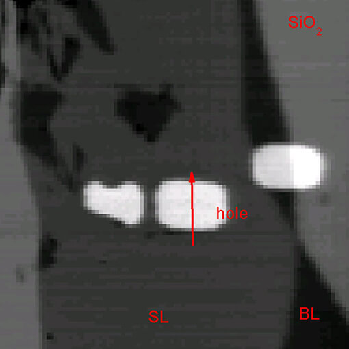
Due to their strong spin-orbit coupling and resulting large spin-orbit splitting in the valence band, layered transition metal dichalcogenides (TMD) are promising materials among the family of 2D materials for spintronic applications. The groups of Nathaniel Stern and Vinayak Dravid at Northwestern University (Evanston, Illinois, USA) used a film of monolayer MoS2 to present a new in-situ electrical biasing technique with transmission electron microscopy. They found that with an applied electric field a net vacancy flux towards the grain boundaries occurs. This vacancy flow results in a regions of Mo-rich nanoparticles aggregating near the voids (see figure c) which appear at strained regions around the grain boundaries (blue colored in figure (b)). The vacancy flow process saturates after several initial biasing cycles when the material reaches a stable state. To characterize these samples, they used the closed cycle cryostat attoDRY2100 combined with the attoCFM I and optical head to study structural dynamics by Raman spectroscopy.
This measurement was realized with the attoCFM I, and the attoDRY2100.
Further reading:
A.A. Murthy et al., ACS Nano 14, 2, 1569-1576 (2020)
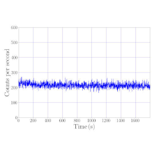
Significant decrease of the optical losses at cryogenic temperature with the attoCFM I
Plasmonic structures present a relevant way to enhance the fluorescence of nanoemitters as colloidal semiconductor nanocrystals. However, they show high optical losses in the visible range which reduce the radiative quantum efficiency. In order to study the evolution of optical losses between 4K and room temperature, the group of Jean-Pierre Hermier at the Université de Versailles Saint-Quentin-en-Yvelines, France, performed time resolved experiments on nanocrystals coupled to a flat gold film. Thanks to the high stability of attoCFM I cooled by a closed cycle cryostat, the attoDRY1100, they recorded the photoluminescence decay at base temperature (4K) of a single CdSe/CdS nanocrystal directly deposited on the flat gold film without any drift for several hours. This is shown by the temporal trace of the intensity in the figure.
As a result of their work and in comparison to the predictions of a theoretical model, the radiative quantum efficiency is enhanced by a factor of three. This motivates for further studies at cryogenic temperatures, especially in the domain of quantum optics for which the reduction of the temperature also results in an improvement of the emitter optical properties.
This measurement was realized with the attoCFM I.
Enhancing Quantum Dot Emitters by Precisely Positioned Micrometric SILs
Using interferometric closed loop scanning integrated into the attoCFM I confocal microscope for cryogenic in-situ lithography, the group of P. Michler in Stuttgart was able to optically localize quantum dots (QDs) with a unprecedented precision of 2 nm and mark them via lithography.
This procedure enables further processing and optimizing these single photon emitters to enhance light extraction. In this case, they successfully demonstrated how to precisely add hemispherical lenses directly on top of the quantum dot via 3D direct laser writing. This led to an enhancement in extraction efficiency by a factor of 2.
“Our attoCFM I LT-lithography setup is not only the best choice when it comes to stability requirements. Its closed loop scanning feature also allows us to optically pre-select quantum dots suitable for desired experiments and mark them in-situ via lithography with nanometric precision.”
Prof. Dr. Peter Michler (University of Stuttgart, Germany)
This measurement was realized with the attoCFM I, and the Low Temperature Photolithography.
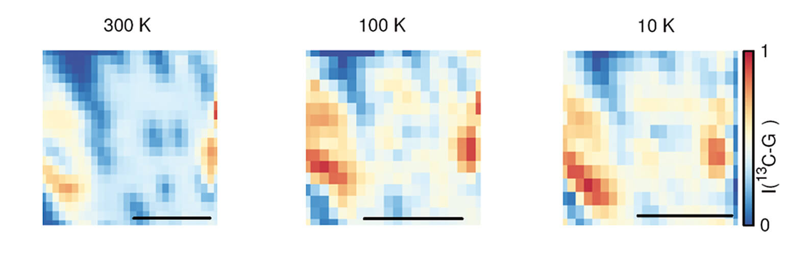
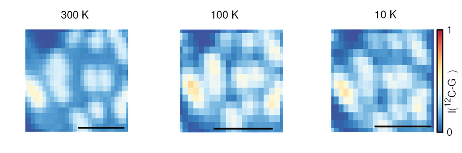
A new way to modulate exciton-complex emissions of TMDs
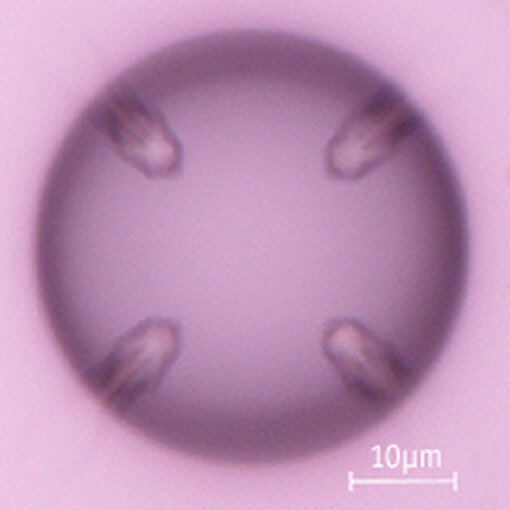
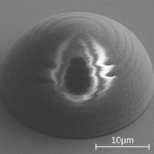
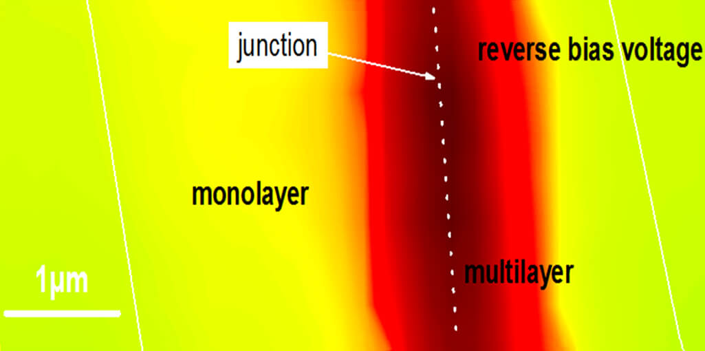
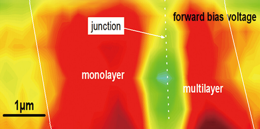
A new type of atomically layered transition-metal dichalcogenides (TMD) was developed by Mr. Taishen Li and co-workers from the University of Science and Technology of China (Hefei, China): a triangular inkslab-like WSe2 homojunction with a monolayer in the inner surrounded by a multilayer frame.Optical and scanning photocurrent microscopy (SPCM) measurements performed with the attoCFM I, cooled by a closed cycle attoDRY1000 to cryogenic temperatures, shows a clear redshift of the photoluminescence peaks from the center to the edge region of the inner monolayer, reflecting a high charge density gradient. In addition, a significant rectifying behavior and photovoltaic response across the homojunction is observed. All in all, the results lead to efficient modulation of the exciton-complex emissions of TMDs.
This measurement was realized with the attoDRY1000, and the attoCFM I.
Further reading:
T. Li, et al., ACS Nano 12 (5), pp 4959–4967 (2018)
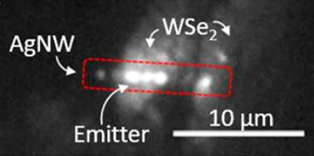
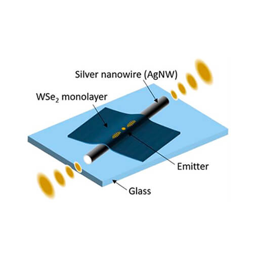
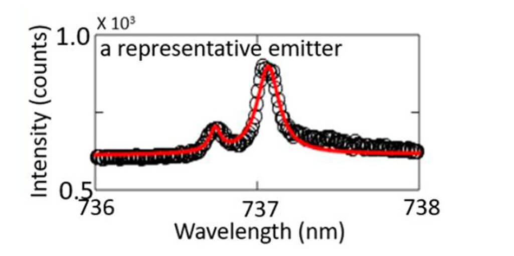
Coupling single defects to a nanowire
Using an attoCFM I cooled by an attoDRY1000, the group of Edo Waks at the University of Maryland (Maryland, USA) succeeded to couple quantum emitters in a tungsten diselenide (WSe²) monolayer self-aligned to the surface plasmon mode of a silver nanowire. The achieved lower bound coupling efficiency was measured to be 26% ± 11% in average from the emitter into the plasmonic mode of the silver nanowire. The presented technique is versatile to construct coupled systems consisting of diverse plasmonic structures and single-defect emitters in a range of two-dimensional semiconductors. Such a coupled system could be used for applications such as ultra-fast single-photon sources, which paves a way toward super-compact plasmonic circuits.
This measurement was realized with the attoDRY1000, and the attoCFM I.
Further reading:
T. Cai, et al., Nano Lett. 17, 6564 (2017)
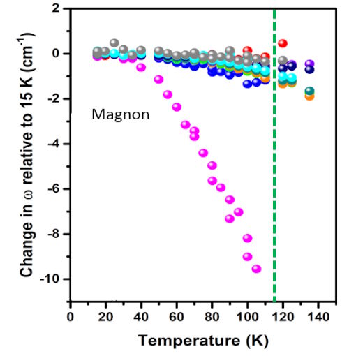
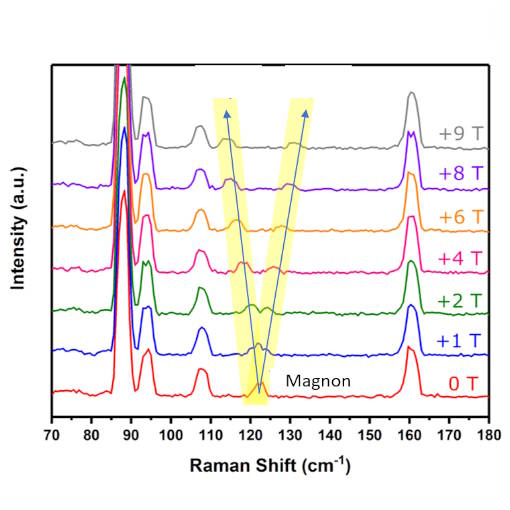
Quasi-2D Magnon identified via Magneto-Raman Spectroscopy
Due to myriad of interesting electronic, mechanical and optical properties, van der Waals materials have been extensively studied in recent years. Exploring their magnetism has been neglected, mainly because of scarcity of long-range magnetic order in 2D materials. Yet, when it exists, like in FePS³, it poses a huge technological potential, particularly since magnetic properties of 2D materials are easily tuneable. The group of Angela Hight Walker at NIST (Gaithersburg, Maryland, USA) developed and used a state-of-the-art cryogenic Raman system cooled by an attoDRY1000 to investigate FePS³ in an antiferromagnetic ordering which leads to a folding of the Brillourin zone and thus to a new modes appearing below the Néel temperature TN. With their temperature and magnetic-field dependent Raman spectra, the group established that one of the appearing modes can only be explained as a magnon and not with a phonon, contrary to previous assumptions. The magnon frequency was detected at 3.7 THz which is an order of magnitude faster than in the previously studied MnPS3. To the best knowledge, this is the first verification of a quasi-2D magnon in a layered material by magneto-Raman spectroscopy.
This measurement was realized with the attoDRY1000, and the attoCFM I.
Further reading:
A. McCreary et al., Phys. Rev. B 101, 064416 (2020)Press release
Scalable Architecture for Multi-Photon Boson Sampling
Research groups led by Jian-Wei Pan & Chao-Yang Lu in China and Sven Höfling in Germany & UK have successfully demonstrated the first quantum simulator based on single photons that beats early classical computers. In Nature Photonics, they report on “High-efficiency multiphoton boson sampling“, implementing 3-, 4-, and 5-boson-sampling with rates which are more than 24,000 times faster than all previous experiments, and 10-100 times faster than the first electronic computer (ENIAC) and transistorized computer (TRADIC) in human history. Their work, which was carefully prepared and accompanied by their 3 previous papers published in PRL (see below), kick starts a new era of photonic quantum technologies-going beyond proof-of-principle demonstrations and building a quantum machine to actually race against different generations of classical computers. In recognition of their achievements in quantum teleportation research, the very active and highly respected Chinese group recently also won the 2015 Physics World Breakthrough of the Year award and the 2015 State Nature Science First Class Award in China. In addition, Chao-Yang Lu was portrayed by Nature last summer as one of the “Science stars of China”. For their quantum dot experiments, his group uses three attoDRY cryostats equipped with attocube positioners, scanners and cryogenic objectives. Visit the group’s homepage for more information on their experiment.
This measurement was realized with the attoDRY1000, and the attoCFM I.
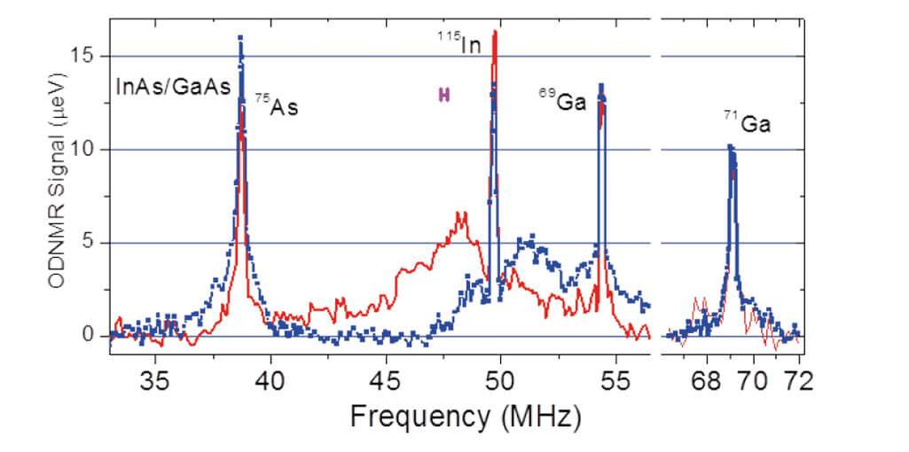
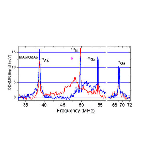
Material Composition and Strain Analysis
Often for material structure analysis, invasive techniques such as e.g. neutron scattering or energy dispersive X-ray spectroscopy inside a focussed ion beam microscope are used to obtain information. Unfortunately, such techniques may generate permanent perturbations to the sample under study.
Researchers at University of Sheffield have developed a modified optically detected Nuclear Magnetic Resonance (ODNMR) technique for low temperature analysis, which allows for structural analysis of strained quantum dots. NMR is a non-invasive technique, which extracts information using the fundamental properties of nucleus for different materials.
The new method is sensitive enough to probe individual strained nanostructures with only 100000 quadrupole nuclear spins. Therefore, this innovative technique proved to be an important non-invasive spectroscopy method for structural analysis especially of strained nanostructures as it requires no special preparation of the sample.
This measurement was realized with the attoCFM I.
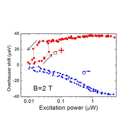
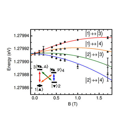
Dynamic nuclear polarisation in GaAs/AlGaAs dots observed at 4 K
In this note, nuclear spin effects are studied in individual quantum dots pumped by circularly polarised excitation using an attoCFM I. Spin is transferred from optically generated electrons to the system of about 10.000 nuclear spins. The effect of nuclear spin polarisation on the electron spin can be described in terms of effective nuclear magnetic field (also referred to as Overhauser field). Optically generated Overhauser fields of the order of several Tesla are observed localised in a single quantum dot of 4 x 20 x 20 nm3 in size.
This measurement was realized with the attoCFM I.
Observation of Many-Body Exciton States using the attoCFM I
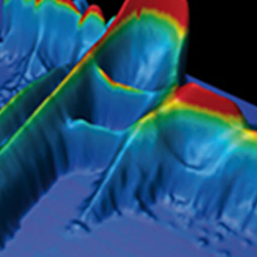
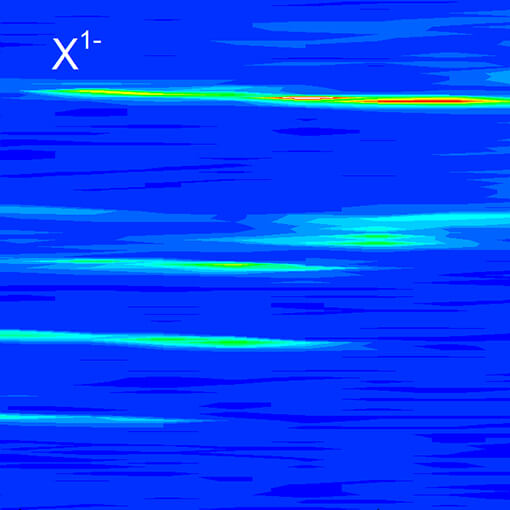
Many-body interactions opens the doors to new fascinating physics such as the Fermi-edge singularity in metals, the Kondo effect in the resistance of metals with magnetic impurities and the fractional quantum Hall effect. The group of Paul M. Koenraad at the Eindhoven University of Technology (Eindhoven, NL) observed striking many-body effects in the optical spectra of a semiconductor quantum dot interacting with a degenerate electron gas using a free beam confocal microscope inside a 3He cryostat.
The image on the left shows a 3D map of the photoluminescence of a single InAs/GaAs quantum dot in a charge-tunable device. It was found that the coupling between the semiconductor quantum dot states and the continuum of the Fermi sea gives rise to new optical transitions, manifesting the formation of many-body exciton states. The experiments are an excellent proof for the stability of the attoCFM as the measurements took more than 15 hours without the need for re-alignment.
This measurement was realized with the attoCFM I.
Further reading:
N. A. J. M. Kleemans et al., Nature Physics 6, 534 - 538 (2010)
Material composition and strain analysis of single semiconductor quantum dots
E.A. Chekhovich and his colleagues at the University of Sheffield developed an elegant modification of an optical technique to analyze material composition in semiconductor quantum dots. This new approach now allows analysis of strained structures, which was extremely difficult before.
This measurement was realized with the attoCFM I.
Low Temperature Raman Measurements on Layers of Graphene
In the attached application note, an attocube attoCFM I optical microscope has been used for high-resolution Magneto-Raman measurements on ultra-pure graphene at low temperatures and high magnetic fields. Anti-crossings can be seen where hybridization of E2g phonons and magneto-excitons take place.
The measurements were performed at 7 K and in magnetic fields up to 9 T in a modified attoCFM I using our new, ultra-stable confocal optics head.
This measurement was realized with the attoCFM I.
Optical absorption on a single semiconductor quantum dot with a magnetic field applied in Voigt geometry
The modular design of the attoCFM III represents a highly flexible and versatile system for high resolution transmission spectroscopy. In these experiments, the setup has been modified to accommodate an inverted geometry. A special confocal objective was designed that redirects the incoming light by 90° to impinge onto the sample perpendicular to the externally applied magnetic field, i.e. in Voigt geometry.
This measurement was realized with the attoCFM I.
Addressing Strain and Doping by Cryogenic Raman Mapping
T. Verhagen in the Czech Academy of Sciences in Prague conducted a comprehensive study on the effects of temperature induced strain on two-layer Graphene sheets using an attoRAMANxs confocal Raman microscope. Using isotopical labelling, they can differentiate the influences of the surface on the lower and on the upper layer. A correlation analysis allows to separate strain and doping contributions to the observed Raman shifts. This detailed analysis allows to estimate temperature induced strain and doping contributions that are important when analyzing transport measurements on graphene mono- and bilayers.
Further reading:
T. G. A. Verhagen, et al., Phys. Rev. B 92 125437 (2015)
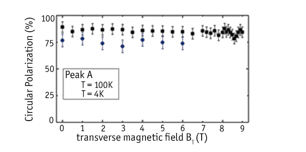
Optical Experiments on MoS2
Once initialized, the electron valley index in momentum space could be used in analogy to the electron charge or spin as an information carrier. Due to its intriguing band structure, this initialization is straightforward in monolayer Molybdenum disulfide (MoS2) an atomically flat, optically active semiconductor. Photoluminescence studies carried out in collaboration between Toulouse University - CNRS (France) and the Chinese Academy of Science provide experimental proof of the robustness of the valley index initialization via polarized laser excitation. The zero field measurements carried out using an attoDry700 table-top cryostat show 90% polarized emission at 4 K with still 40% of polarization remaining at room temperature. Measurements in an attoDRY1000 magneto-cryostat in a transverse magnetic field up to 9 T at 4 K (6 T at 100 K) confirm the robust electron valley index initialization.
(Images courtesy of Bernhard Urbaszek and co-workers.)
This measurement was realized with the attoCFM I.
Further reading:
G. Sallen, et al., Phys. Rev. B 86, 081301(R) (2012)
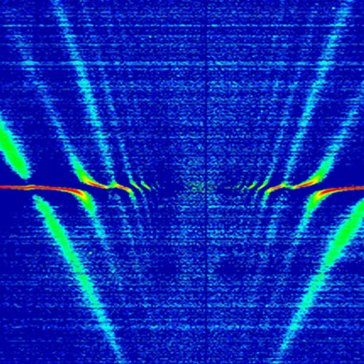
Raman Spectroscopy on Graphene
The figure to the left shows magneto-Raman measurements recorded at 4 K on an exfoliated single crystal of natural graphite with unprecedented spatial resolution (approx. 0.5 µm), while sweeping the magnetic field from -9 T to +9 T. The data were recorded on a single graphene flake and demonstrate the crossing of the E2g phonon energy with the electron-hole separation between the valence and conduction Landau levels.
(-N,+M) of the Dirac cone. Resonant hybridization of the E2g phonon is a specific signature of graphene flakes which display very rich Raman scattering spectra varying strongly as function of magnetic field [1].
[1] C. Faugeras et al., Phys. Rev. Lett. 107, 036807 (2011);
(attocube application labs, 2011; work in cooperation with C. Faugeras, P. Kossacki, and M. Potemski, LNCM I - Grenoble, CNRS_UJF_UPS_INSA France)
This measurement was realized with the attoCFM I.
Further reading:
Raman Spectroscopy on Graphene made with attoRAMAN
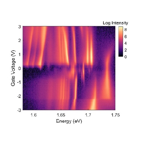
Resonant Spectroscopy on a Single QD
Spectroscopy of semiconductor quantum dots (QDs) under resonant optical laser excitation and of other single photon emitters, such as vacancy-centers often yields more information about the emitters than more ubiquitous non-resonant excitation. However, it is a technically challenging measurement to perform. The difficulty lies within the separation of the excitation laser photons from the re-emitted and scattered photons. One way in which this can be achieved is by means of polarization suppression: in a geometry where the scattered laser photons have a well-defined polarization, they can be filtered from the detected signal facilitating the detection of resonance fluorescence (RF) of a single quantum dot or any other quantum emitter.
The attoCFM I can be upgraded with a resonant fluorescence package, which features an apochromatic performance that permits alignment free switching between off resonant PL measurements and RF. This feature is fully enabled by our novel cryogenic compatible apochromatic objectives designed to hold the focus plane at the same position on the sample independently from the photon wavelength.
The combination of high precision rotators with the flexible beam management of the confocal CFM I head leads to an easy and reproducible use for our customer. It provides extinction ratios of 107, just a factor 10 away from the world record in research labs while allowing an unprecedented flexibility of use.
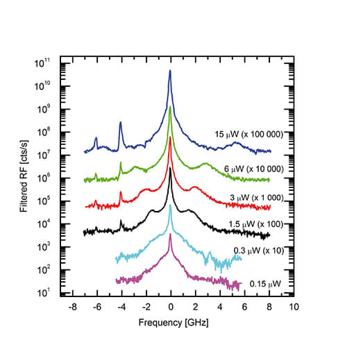
The first figure shows the resonance fluorescence of a quantum dot measured with the attoCFM I equipped with the Polarization Extinction Option and a narrow band tunable laser. In order to resolve the Mollow triplet, the emission is filtered through a high resolution spectrometer. Here, the extinction ratio exceeds 106, using the low temperature near infrared apochromatic objective LT-APO\NIR.
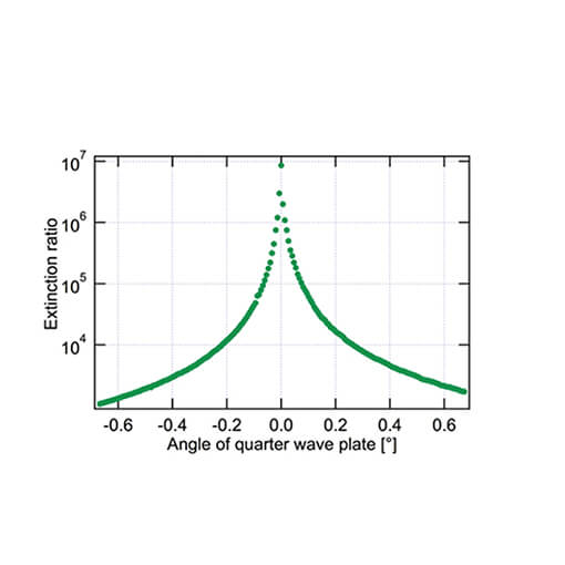
The second figure shows the extinction ratio of the Polarization Extinction option for the attoCFM I as a function of the rotation angle of the inbuilt piezo rotator equipped with a quarter wave plate. In an angular region of 30 m° an extinction of more than 106 can be reached with a tunable narrow band diode laser (<1?pm line width).
Measurement and data by E. Kammann (1), S.H. E. Müller (1), K. Puschkarsky (2), M. Hauck (2), S. Beavan (2), A. Högele (2), and K. Karrai (1),
(1) attocube systems AG, Munich, Germany,
(2) Ludwig Maximilian Universität, Munich, Germany
This measurement was realized with the attoCFM I.
Resonance Fluorescence Spectroscopy
In this Application Note, we present resonance fluorescence spectroscopy measurements on a semiconductor quantum dot measured with our attoCFM I. We successfully employed the new polarization extinction upgrade in combination with our new infrared ranged, apochromatic performance, low-temperature objectives. Spectral filtering of the emission reveals the Mollow triplet.
This measurement was realized with the attoCFM I.
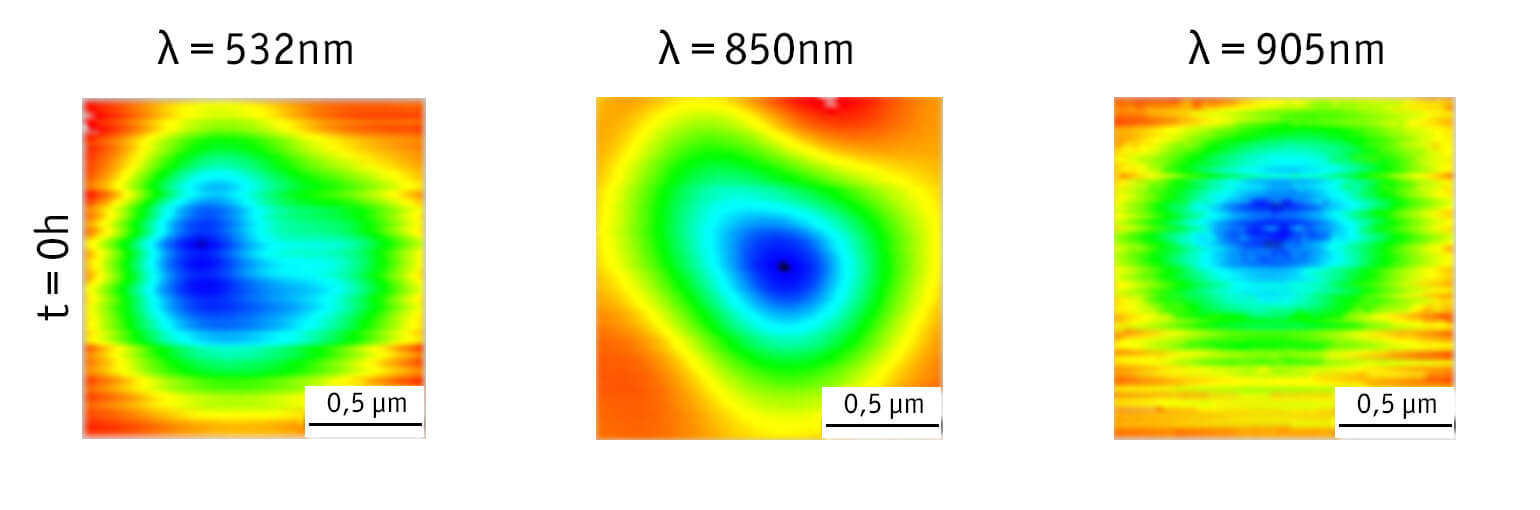
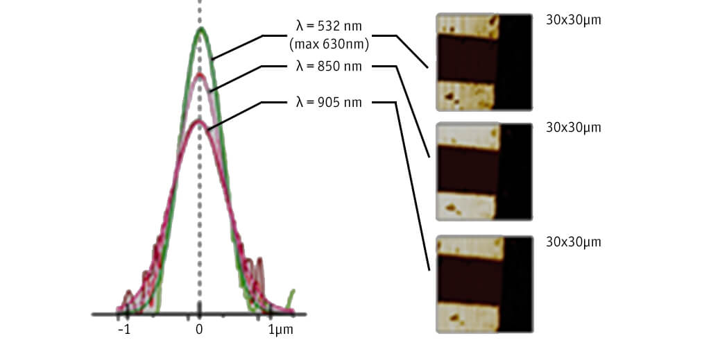
3 color spot high resolution alignment
The figure to the left shows a superposition of the focused spots of three CFM channels at 4 K with the following key features:
- Spots alignment resolution ±15 nm
- Absolute positioning (contouring) error: 12 nm @ 1 µm/s
- Long term drift less than 10 nm/h
(attocube application labs, 2011)
This measurement was realized with the attoCFM I.
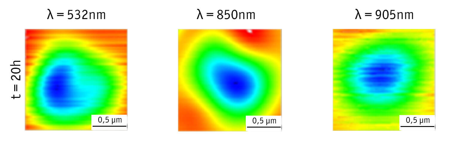
Remarkable Long Term Stability
Long term drift measurements recorded at cryogenic temperatures on a single quantum object using all three channels of the attoCFM. The position drifts during 20 hours of measurement time account for less than ±10 nm/hr in X and Y direction. Image size is 1.25 x 1.25 µm2.
(attocube application labs, 2011)
This measurement was realized with the attoCFM I.