
Nanoparticle Imprinted Matrices
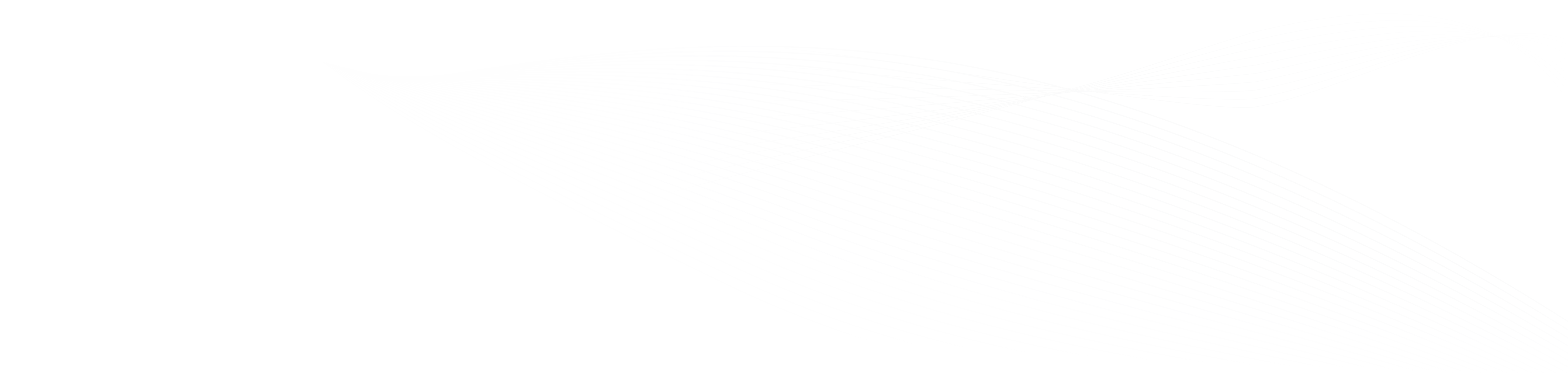
Nanoparticle-imprinted matrix is an innovative approach that allows for the selective detection of nanoparticles based on their size, shape, surface chemistry, and composition. However, those systems have mainly been used for detecting metallic nanoparticles such as Au or Ag by electrochemical dissolution. This study applies s-SNOM imaging and AFM-IR spectroscopy to characterize the physical entrapment of silica-NPs by the matrix at the nanoscale. Point IR spectra collected from a 20 nm radius identified the vibrations of the aromatic C=C bond and the amine group of the poly(3-aminophenol) matrix, as well as the Si-O-Si vibration of the silica nanoparticles. In addition, IR-sSNOM imaging at specific wavenumbers demonstrated the intricate dispersion of silica-NPs enclosed by the matrix and revealed the chemical and vibrational contrast in the nanoscale between the organic matrix and the inorganic nanoparticles.In summary, the use of AFM-IR and s-SNOM technologies in this study provided unique insights into the electrochemical reduction of CO2 to CO and demonstrated the power of these advanced analytical techniques for the investigation of complex chemical reactions.
This measurement was realized with the IR-neaSCOPE.
Further reading:
Dery et al., ChemElectroChem e202300039, (2023)
Lamella-forming PS-b-PMMA Films
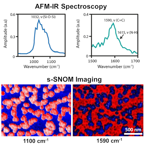
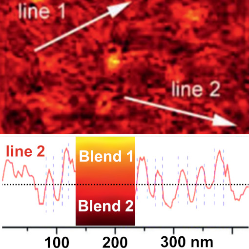
A non-invasive, image-based analytic method utilizing s-SNOM is suggested to evaluate the phase separation behavior of lamella-forming PS-b-PMMA block copolymer films. Taking advantage of the penetrability of the tip-enhanced IR signal into the films, the spatio-spectral maps of each component are constructed. Subsequently, the effect of a sole and combinatorial applications of the self-assembly procedures, such as solvent vapour annealing (SVA) and/or thermal annealing (TA), on the spatial distribution of PS or PMMA components is quantitatively assessed in terms of the areal portions of the PS domain, PMMA domain, and the mixed zone that is adjacent to the domain border. Additionally, by statistically comparing the local concentration profiles, the chemical contrast between the domains turns out to be dependent upon the annealing procedures (namely, SVA and SVA+TA).
s-SNOM technique can pave the way to an uncomplicated but precise investigation of the polymer nanostructure-based thin film devices, whose performances are critically governed by the spatial arrangement of the chemical elements.
This measurement was realized with the IR-neaSCOPE.
Further reading: Kim et al., Spectrochimica Acta Part A 274, 121095 (2022)
High-efficient Organic Photovoltaics
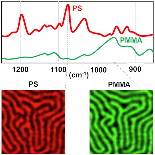
In organic photovoltaics, morphological control of donor and acceptor domains on the nanoscale is the key for enabling efficient exciton diffusion and dissociation, carrier transport and suppression of recombination losses. This publication demonstrates a double-fibril network based on a ternary donor–acceptor morphology with multi-length scales constructed by combining ancillary conjugated polymer crystallizers and a non-fullerene acceptor filament assembly. Essential for this study was the nanoscale infrared image of double-fibril network PM6/L8-BO with strong IR contrast at 1648/1532 cm-1. In addition, the line profiles across the image was used to estimate a fibril diameter of 22.1 and 22.6 nm for acceptor and donor fibrils concluding that the volume of the mixing domain is low, resulting in low geminate recombination and a high fill factor.
To summarize, double-fibril network morphology strategy minimizes losses and maximizes the power output, offering the possibility of 20% power conversion efficiencies in single-junction organic photovoltaics.
This measurement was realized with the IR-neaSCOPE.
Further reading:
Zhu et al., nature materials 21, 656 (2022)
E-Book on Polymer Nanostructures
Nanocomposite polymers, multilayer thin films, nanofibers and other polymer nanoforms often offer new properties or enhanced performance compared to bulk materials, demanding tools for chemical analysis with nanoscale spatial resolution for their investigations. We introduce in this e-book leading techniques for nanoscale chemical mapping and identification.
Applications covered in this e-book:
- Nanoscale chemical identification using standard IR database,
- IR nanoimaging of polymer films, nanoparticles & monolayers,
- Molecular conformation and orientation in an ultrathin polymer film,
- Hyperspectral & correlative nanoscopy of polymer composites.
This measurement was realized with the IR-neaSCOPE.
Further reading:
E-Book Download
Flexible Organic Photovoltaics
Organic photovoltaics hold great potential for cost-effective, sustainable and flexible devices production. On the other side, organic semiconductor films are semicrystalline material with energetic disorder, inefficient carrier transport and high energy loss. Several studies focused on morphology strategy proposed the development of better suited phase separation to improved structure order, facilitate efficient transport and suppress trap states. This report uses tapping AFM-IR+ to map mixtures of organic photovoltaic blends and fine-tune the material morphology and crystallization. The Authors concludes that mixtures of blends with structural similarity but with different electronic properties exhibit significant improvement of high-crystallinity, fibrillar morphology and even shows enhancement in performance.
In contrast to electrons, IR photons extends the structural investigation beyond the top atomic layer complementing current nanoscale investigation methodology. Therefore, the IR-based investigation methods (AFM-IR, s-SNOM and nano-FTIR) can be also applied to other thin-film materials, e.g. sensors, electronic equipments or semiconductor devices.
This measurement was realized with the IR-neaSCOPE.
Further reading:
Zhang et al., Advanced Materials 33, 2007177 (2021)
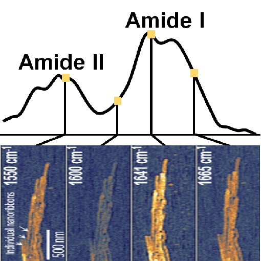
Mineralization of Protein Nanoribbons
Dental enamel is the hardest tissue created by vertebrates and exhibits complex nanoscale organizations of apatite crystals and protein nanoribbons. Previous studies using AFM and TEM show the formation of nanoribbons and even suggested their twisted structure. In this study, tapping AFM-IR+ provides additional chemically-specific information by measuring local infrared sample absorption. And indeed, absorption in the amide I and amide II bands verifies the protein composition of nanoribbons and provides insights into their α-helix/β-turn secondary structure. In addition to basic understanding of enamel growth, this study it is an important step towards synthesis of nanostructured materials using self-assembled organic framework.
Using low energy photons, tapping AFM-IR+ is highly suitable for nanoscale imaging and spectroscopy of fragile biological samples, such as protein, fibrils, lipids, viruses and DNA.
This measurement was realized with the IR-neaSCOPE.
Further reading:
Bai et al., PNAS 117, 19201-19208 (2020)